Case 1
A 28-year-old man presented with a left testicular mass. Tumour markers were taken and he went on to have a radical inguinal orchidectomy. The specimen and histology are shown.


- Which testicular tumour markers were taken?
- What does their elevation signify?
- What is their half-life?
- Describe the histology shown?
- What features define high-risk disease for stage 1 seminoma and non-seminomatous germ cell tumour (GCT)?
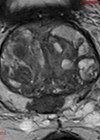
Case 2
A 65-year-old female underwent a left laparoscopic nephrectomy for a renal mass. The histology is shown (low and high powered microscopy).


- Describe the key histological features? What is the diagnosis?
- What grading system can be applied to these tumours?
- Define this grading system.
Case 3
An 84-year-old man underwent a cystoscopy and bladder biopsy for a red patch in his bladder. The images and histology are shown.


- What is the diagnosis? Explain the histological features.
- With no subsequent treatment, what proportions of these patients develop muscle invasive disease?
- What are the benefits of treating with intravesical BCG?
- How are BCG failures managed?
Uropathology: what’s the diagnosis? – Answers
Case 1
-
α-fetoprotein, β-human chorionic gonadotropin (β-HCG), lactate dehydrogenase (LDH).
-
α-fetoprotein: non-seminoma GCT, hepatic dysfunction, hepatitis, cirrhosis. β-HCG: either seminoma or non-seminoma GCT, trophoblastic differentiation in lung / gastric tumours, marijuana use. LDH: non-specific tumour marker reflecting tumour burden, growth rate and cellular proliferation.
-
α-fetoprotein: five to seven days, β-HCG: two to three days.
-
Macroscopically: 1.5cm solid, yellow mass in the rete testes, confined to the testes. Microscopically: Normal testis on the left side. There are lobules of neoplastic cells with intervening stroma with characteristic lymphoid infiltrates. These are classical seminoma cells, with large vesicular nuclei and pale watery cytoplasm.
-
Seminoma: Tumour size >4cm and rete testes invasion. Non-seminomatous tumours: vascular invasion.
Case 2
-
The lesion on the right hand side shows cells with clear cytoplasms, which are arranged in nests. Nuclear atypia is seen on higher magnification. These are consistent with clear cell renal carcinoma.
-
Fuhrman nuclear grade – based around nuclear appearance.
-
Fuhrman grade is based on the highest grade present if heterogeneous, with a scale from 1-4. Grade 1: round and uniform nuclei. Grade 2: slightly irregular nuclear contours. Grade 3: moderate to markedly irregular nuclear contents. Grade 4: multilobular nuclei, large / prominent nucleoli.
Case 3
-
Carcinoma in-situ. Atypical hyperchromatic cells with significant variation in nuclear size and shape. The lamina propria is hypervascular with scattered inflammatory cells.
-
Fifty-four percent will progress to muscle invasive disease.
-
Complete response rate of 72-93% with BCG, plus reduced risk of progression to muscle invasive disease by 35%.
-
Non-high grade recurrence after BCG is not considered as BCG failure. The evidence-based treatment is radical cystectomy, with other treatments being oncologically inferior.